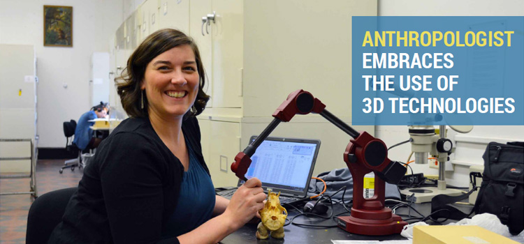
As a biological anthropologist, Claire Terhune spends a majority of her time studying the skulls of living primates, modern humans, and human ancestors.
Working with a team of undergraduate and graduate students in the Department of Anthropology at the University of Arkansas, her lab is dedicated to understanding how modern humans evolved from other primates, and investigating how early humans migrated out of Africa and dispersed into Asia and Europe.
Skull Holds the Key to Understanding Our Past
The skull’s shape, size, and characteristics contain a wealth of knowledge about a specimen’s genetics, environment, evolutionary changes, dietary preference, and how it once lived. For instance, by comparing the chewing apparatus and dentition of the modern human skull to our fossil ancestors, Terhune discovers how diet and feeding behaviors have evolved over the past ~7 million years.
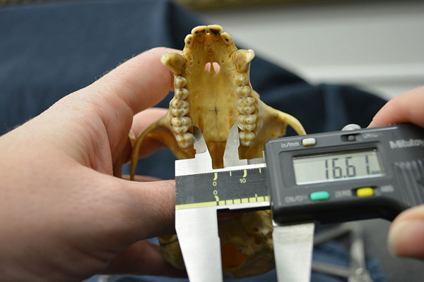
Isolating specific characteristics and features of various skulls for comparative analysis can be a difficult endeavor. The skull is a complex three-dimensional (3D) biological structure. While calipers are good at taking simple measurements such as length or width, collecting large amounts of data using this measurement device can be time consuming. They are limited in the types of measurements that can be collected and analyses that can be performed.
Using 3D Scanning as an Advanced Quantitative Technique
“Biological anthropologists are rapidly embracing the use of 3D technologies in their research,” Terhune says. “Advanced quantitative techniques such as 3D scanning help us capture complex shape variations and they form a major component of the work being undertaken in our lab.”
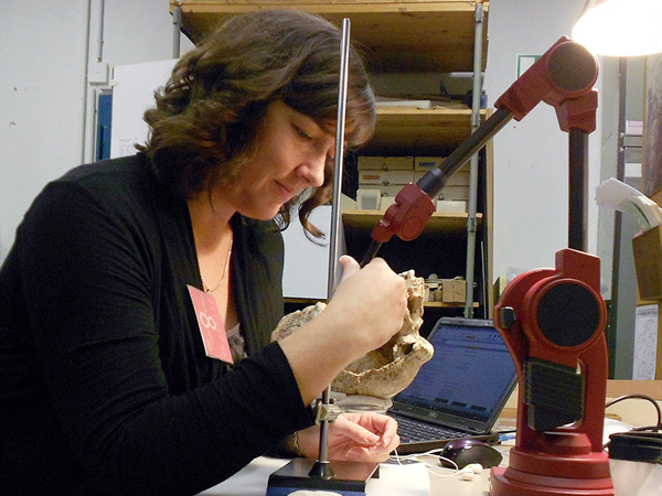
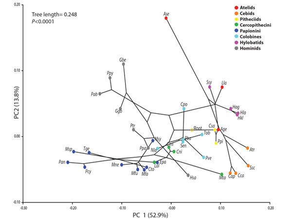
Terhune has been using the MicroScribe 3D digitizer for more than 10 years, particularly for landmark studies using 3D geometric morphometric techniques.1 When she joined the University of Arkansas in 2014, she purchased the MicroScribe portable CMM from GoMeasure3D as an addition for the lab.
“My research takes me to different places all over the world,” Terhune explains. “The MicroScribe 3D digitizer is a portable solution I can easily take with me on my travels. I can quickly set it up at the museum I’m visiting and capture the X,Y, Z landmarks (3D geometric morphometric data) I need just by touching the skull with the articulating arm.”
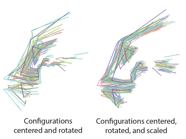
With the data collected, she compares these coordinates across different specimens to see how they relate to or differ from one another. Terhune has published a series of research papers using MicroScribe data that formed the basis of her research findings.
Adding a Structured-Light 3D Scanner for the Lab
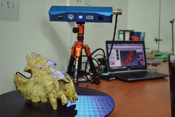
Terhune was looking to add a structured-light 3D scanner for her lab and asked GoMeasure3D for assistance in finding suitable equipment that would meet her needs. The primary purpose was to find a 3D scanner that would digitize a wide variety of skulls and fossils in their entirety and with high levels of accuracy.
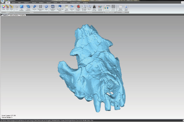
Typically researchers in her team have a limited amount of time with the specimen they are examining, whether they are in a remote location or visiting a museum. With the use of a 3D scanner, they can capture a full digital 3D model of the specimen containing all of the surface measurements. This allows them to ensure nothing was missed during the data collection process, and her team can then reference the digital file for measurements or further analysis at any time.
“If we have a fossil that is crushed, we can use the HDI 3D scanner to recreate it in 3D by scanning the fragments.
Technology enables us to piece prehistory back together and gives us the opportunity to learn more about our past.”
Claire Terhune Assistant Professor, Department of Anthropology University of Arkansas
“Claire was looking for a 3D scanner with great accuracy, high resolution, and portability,” Darryl Motley of GoMeasure3D explains. “Her colleagues were using 3D scanners costing upwards of $50,000-60,000 and she was looking for a more affordable solution. Systems costing a few thousand dollars didn’t give her the quality she was looking for. We found the HDI 3D scanner was the best solution for her in terms of the quality it provides while spending a fraction of the cost of a very high-end 3D scanner.”
Since acquiring the HDI 3D scanner, Terhune has been using it for landmark studies, documentation, and fossil reconstruction. GoMeasure3D also recommended getting a motorized turntable with the HDI 3D scanner to automate the 3D scanning process. “The turntable is a real time saver for us as it eliminates most of the manual work of stitching individual scans together in the software”, Terhune explains. “We can focus more time analyzing the data rather than collecting it.”
Romania Research Trip
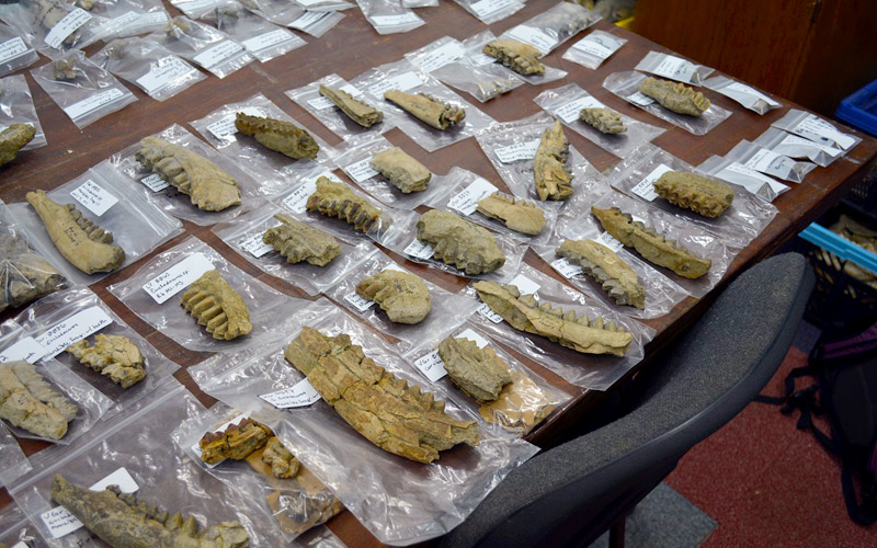
Terhune took the HDI 3D scanner with her on a recent research trip to Romania. She and several colleagues2 went to Bucharest, Romania, to catalog, analyze, and digitize fossils from Grănceanu, Romania, a paleontological site between 1.5 to 2 million years old. They examined these fossils in detail to find evidence that may explain how our human ancestors first migrated out from Africa and eventually dispersed throughout Europe.
They plan to publish their preliminary findings using the data acquired from the HDI 3D scanner later this year. The National Science Foundation (NSF) also awarded the team a $30,000 grant to conduct fossil surveys in Olteţ River Valley of Romania in the summer of 2017. Terhune’s colleague has a HDI 3D scanner as well and they will be using both scanners to maximize their scanning workflow.
“We can’t simply rely on using only one measurement for our research,” Terhune explains. “That’s why we have a range of measurement tools in our lab from calipers, a MicroScribe 3D digitizer, to the HDI 3D scanner. I like using multiple measurement techniques depending on what I need. For example, there’s no point in 3D scanning a complete skull if you just need one quick measurement, but if we want to capture the overall shape and geometry of a complex object, 3D scanning is the way to go. To make sure we have maximum flexibility, we need a data collection ‘toolbox’ with a comprehensive set of measurement tools so we can use the right tool that’s up for the task.”
Next Steps
Terhune and her colleagues3 recently received a $219,000 grant from the NSF to study skull and jaw anatomy of 16 closely related primate species, including humans. They will examine how the teeth, jaw and related joints work together as a chewing system and how they are affected by aging and abnormal changes. Part of the research involves traveling to different museums to scan skulls using the HDI 3D scanner to collect data for statistical analysis. When their research is concluded, these models will be made available on data sharing websites such as MorphoSource.
“Once we’ve collected all of the data, we will make it available to other researchers”, Terhune says. “Specifically, we want to make it easier for junior researchers or those without the ability to travel to museum collections to access and use these data in their research, and decrease the wear and tear on museum collections. Most importantly, through collaboration and building on each other’s research, we can advance anthropological research in our field by discovering more about our shared evolutionary path.”
– – –
About Claire Terhune
Claire Terhune is an assistant professor in the Department of Anthropology at the University of Arkansas. As a biological anthropologist, her research focuses on evaluating human and primate variation in an evolutionary and biomechanical framework. She is especially interested in understanding what makes humans unique. For more information, please visit the Terhune Lab.
– – –
References:
- Geometric morphometrics is the comparative study of complex biological structures by referencing a set of common anatomical landmark points (3D coordinates) across different specimens.
- Sabrina Curran (Ohio University), Chris Robinson (Bronx Community College, City University of New York), and Marius Robu and Alex Petculescu, both with the “Emil Racoviţă” Institute of Speleology
- Siobhan Cooke (Johns Hopkins School of Medicine) and Claire Kirchhoff (Marquette University)